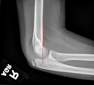
The pediatric elbow - most non-pediatric radiologists strongly dislike the pediatric elbow as there are many things going on. In elbows, adult and pediatric alike, looking at the alignment and for elevated fat pads is always the first step. In this case, you can see that both of the fat pads are elevated, which indicates an underlying effusion from hemarthrosis. So, in the setting of trauma, we at LEAST know there is an occult fracture present.
 |
| Positive Sail Sign - Hemarthrosis |
In adults, this most commonly indicates a radial head fracture, but in children, it is most commonly a supracondylar fracture. At this point, looking at the alignment allows you to evaluate for a supracondylar fracture using the anterior humeral line. The anterior humeral line should cross through the middle third of the capitellum. In this case, you can see that the anterior humeral line is normal.
In pediatric elbows, evaluating the ossification centers using the mnemonic, CRITOE is important. This mnemonic stands for the order of formation/appearance of the ossification centers in the elbow and the approximate age at which they appear (ages 1, 3, 5, 7, 9, 11).
Capitellum - age 1
Radial head - age 3
Internal (Medial) epicondyle - age 5
Trochlea - age 7
Olecranon - age 9
External (Lateral) epicondyle - age 11
In our case, we have a 9 year old, so we should have all of the ossification centers present, except the lateral epicondyle, and they should be normally positioned. Our patient has all present (including the external/lateral epicondyle) and all are normally positioned. Sometimes, after all that work, pediatric elbows are not fulfilling because you can't always find the fracture. At this point, I would dictate this case as positive for elbow joint effusion, consistent with occult fracture.
Here is a good example of a supracondylar fracture:
 |
| AHL - through the anterior third of the capitellum |
Pediatric Elbow


